Home » Products » Scanning Electron Microscopy » Scanning Electron Microscopy - SEM » Tescan Life Science SEM
Tescan Life Science SEM
|
TEScan Scanning Electron Microscopes for Life Science
Tescan Life Science SEM
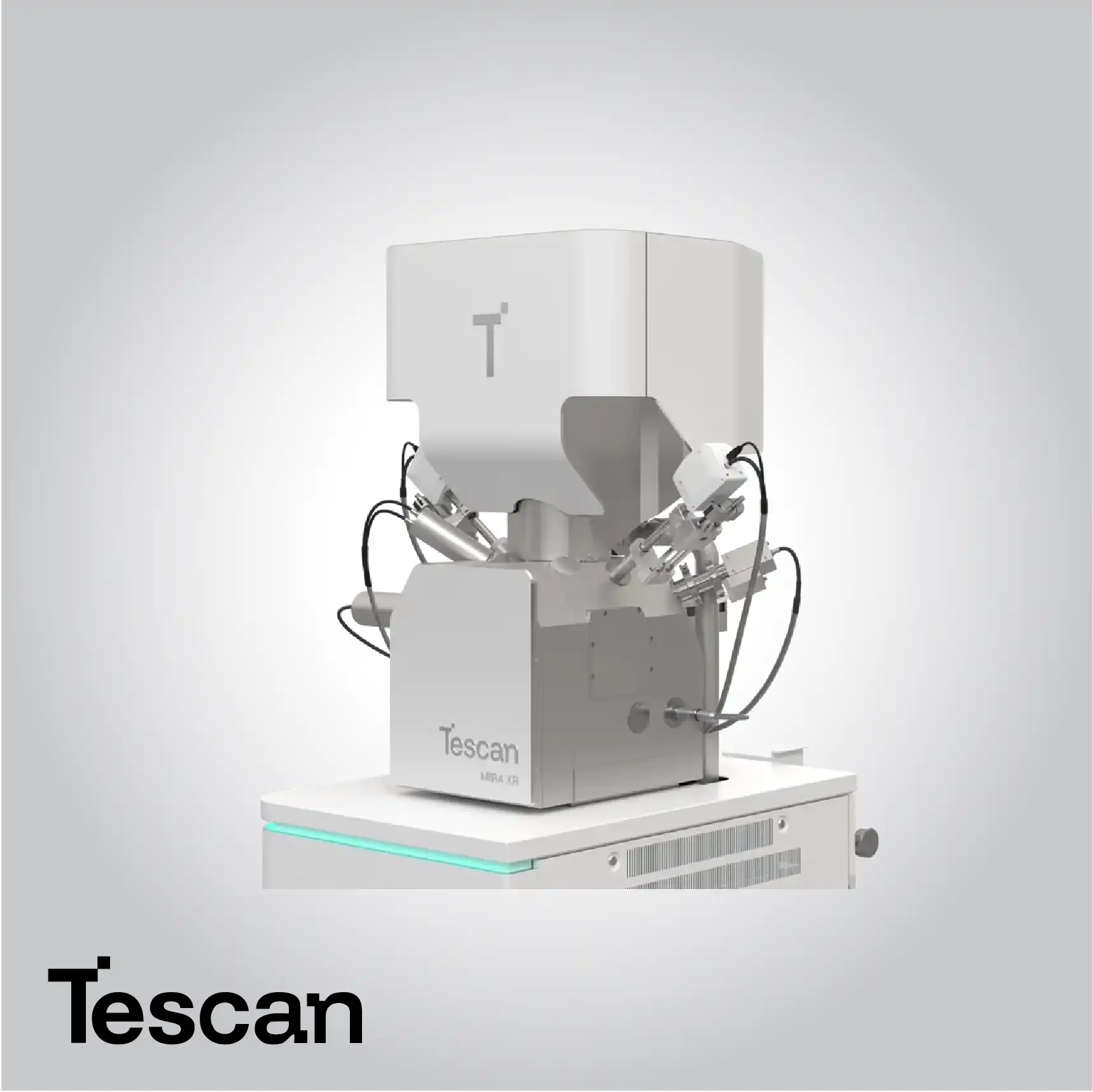
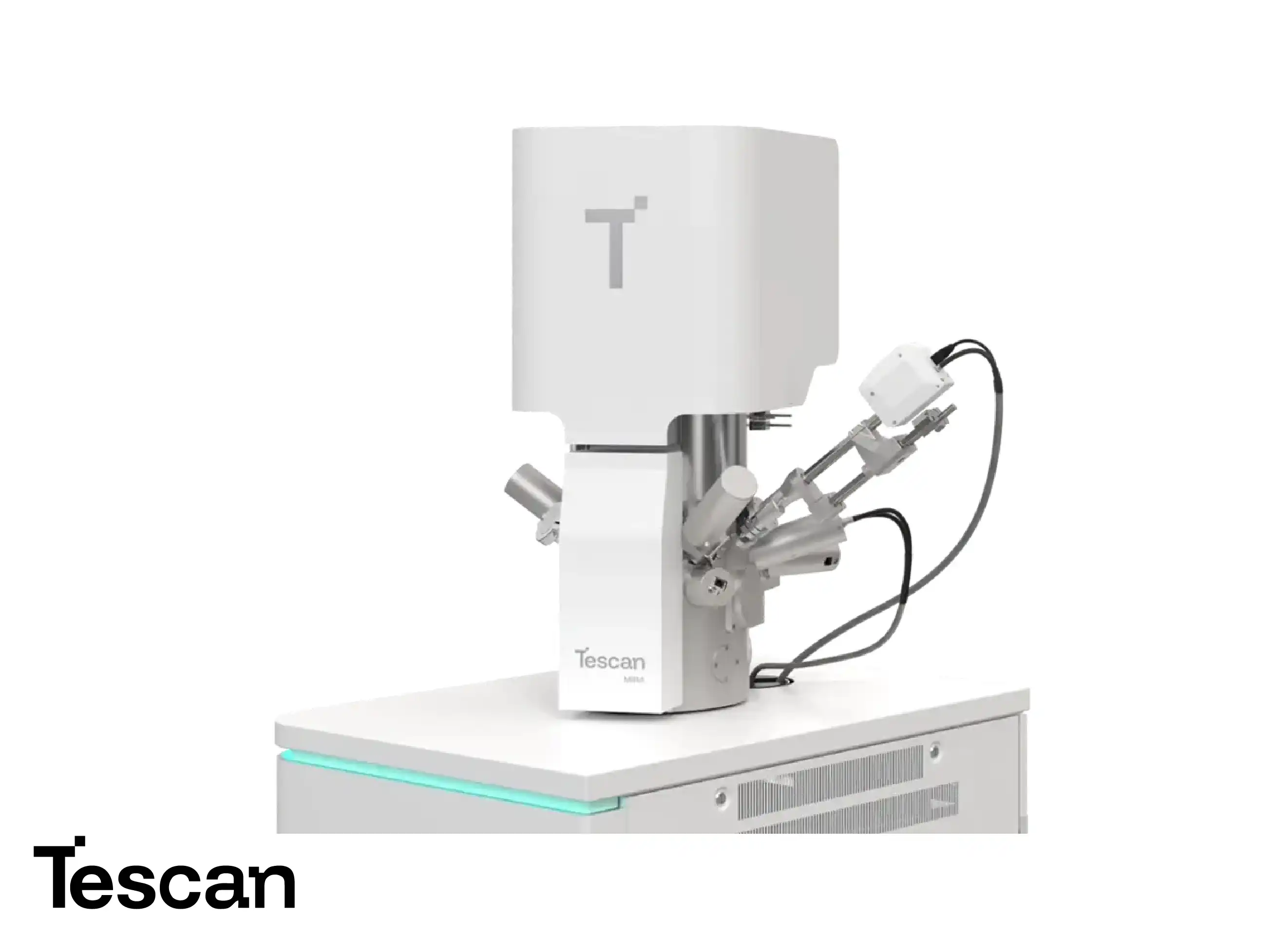
TEScan Life Science Scanning Electron Microscopes provide exceptional imaging and analytical capabilities for biological and biomedical research. These SEM systems enable high-resolution visualization of cells, tissues, and microorganisms, delivering detailed insights into structural organization and morphology. With minimal sample preparation, researchers can preserve delicate biological samples while achieving precise and reproducible imaging results.
TEScan SEM systems for life science are equipped with advanced electron beam control and sensitive detectors, allowing the study of surface structures, fine details, and subcellular components. These systems support both environmental and high-vacuum imaging modes, providing flexibility for a wide range of biological specimens while maintaining optimal image quality.
High-Resolution Biological Imaging
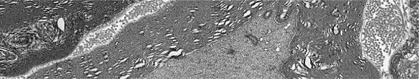
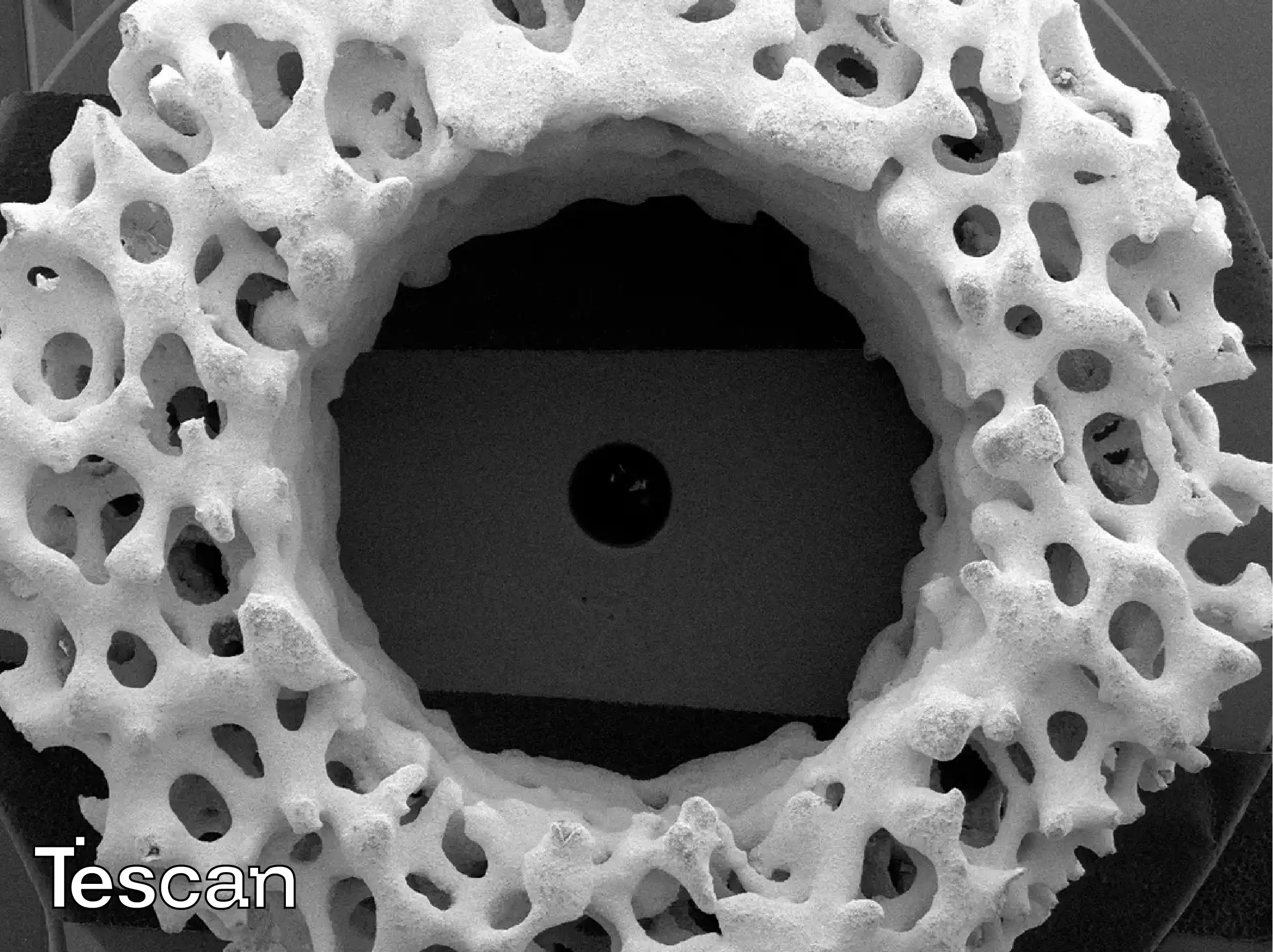
TEScan SEM systems enable researchers to visualize biological structures down to the submicron level, capturing complex surface morphologies and ultrastructural details with outstanding clarity. The optimized electron beam column ensures exceptional image sharpness, allowing accurate differentiation between cellular membranes, extracellular matrices, and subcellular components. This level of precision helps researchers identify morphological variations related to biological function and pathology, supporting both fundamental and applied life science investigations.
Environmental and High-Vacuum Imaging
TEScan SEM instruments feature both environmental and high-vacuum imaging capabilities, allowing researchers to investigate hydrated or uncoated biological samples without the need for extensive sample preparation. Environmental modes maintain controlled pressure and humidity around the specimen, minimizing dehydration and structural collapse. This flexibility enables observation of biological samples in more natural states, bridging the gap between conventional electron microscopy and in-situ biological research.
Structural and Morphological Analysis
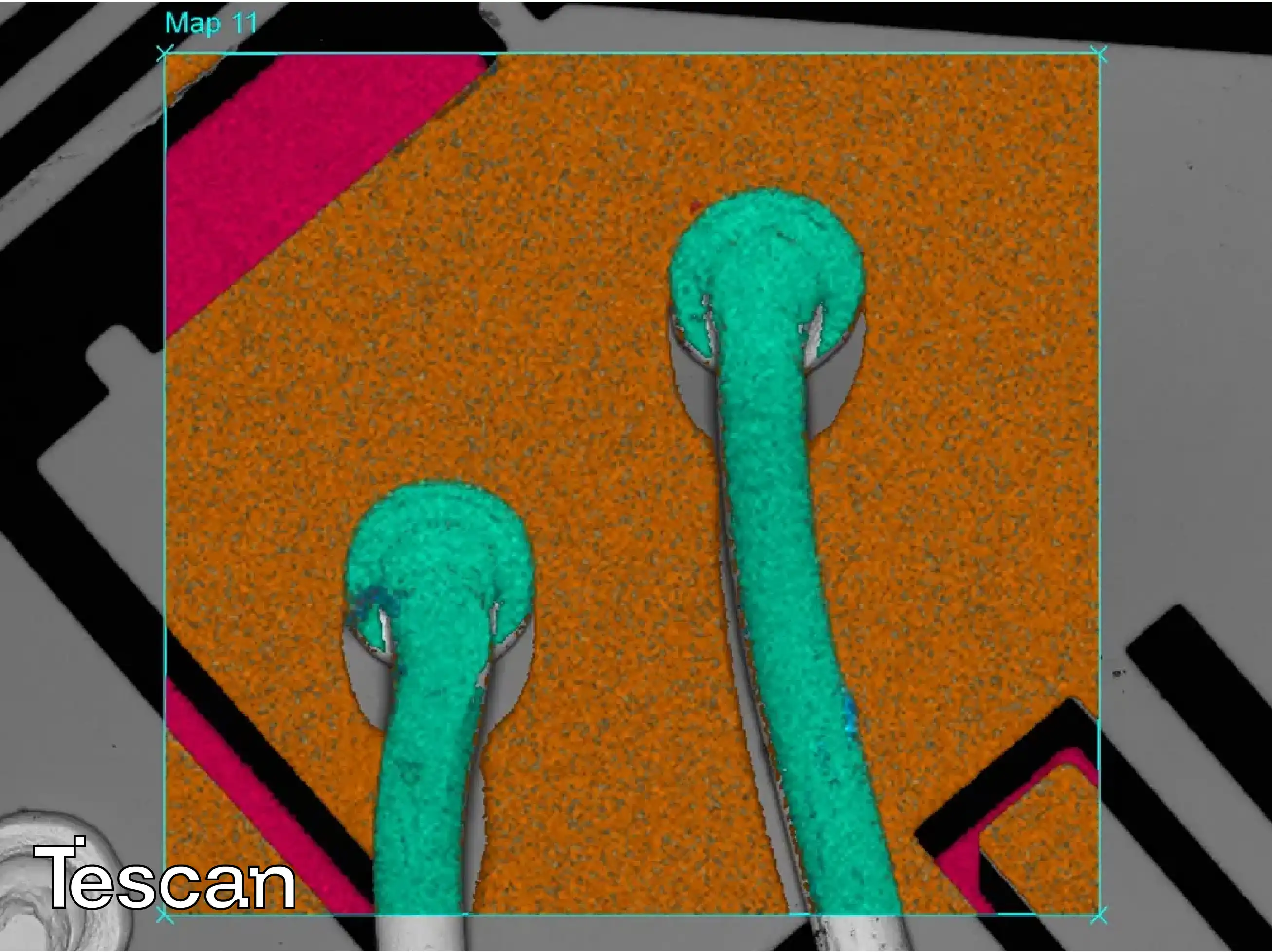
TEScan SEM allows detailed characterization of biological microstructures, enabling analysis of cellular organization, tissue texture, and bio-material interfaces. High-contrast imaging reveals structural patterns, while analytical attachments can provide elemental mapping for compositional insight. This combination of structural and chemical information supports multidisciplinary research, from studying cellular development to understanding the behavior of bioengineered materials.
Advanced Electron Optics and Beam Control
The electron optics architecture of TEScan SEM ensures stable beam performance and minimal drift, even at low accelerating voltages required for biological samples. Precision beam alignment and optimized scanning parameters result in uniform illumination and reduced charging effects, producing images with consistent contrast and resolution. This allows for prolonged imaging sessions and accurate data acquisition from sensitive biological materials.
Detector Innovation for Life Science
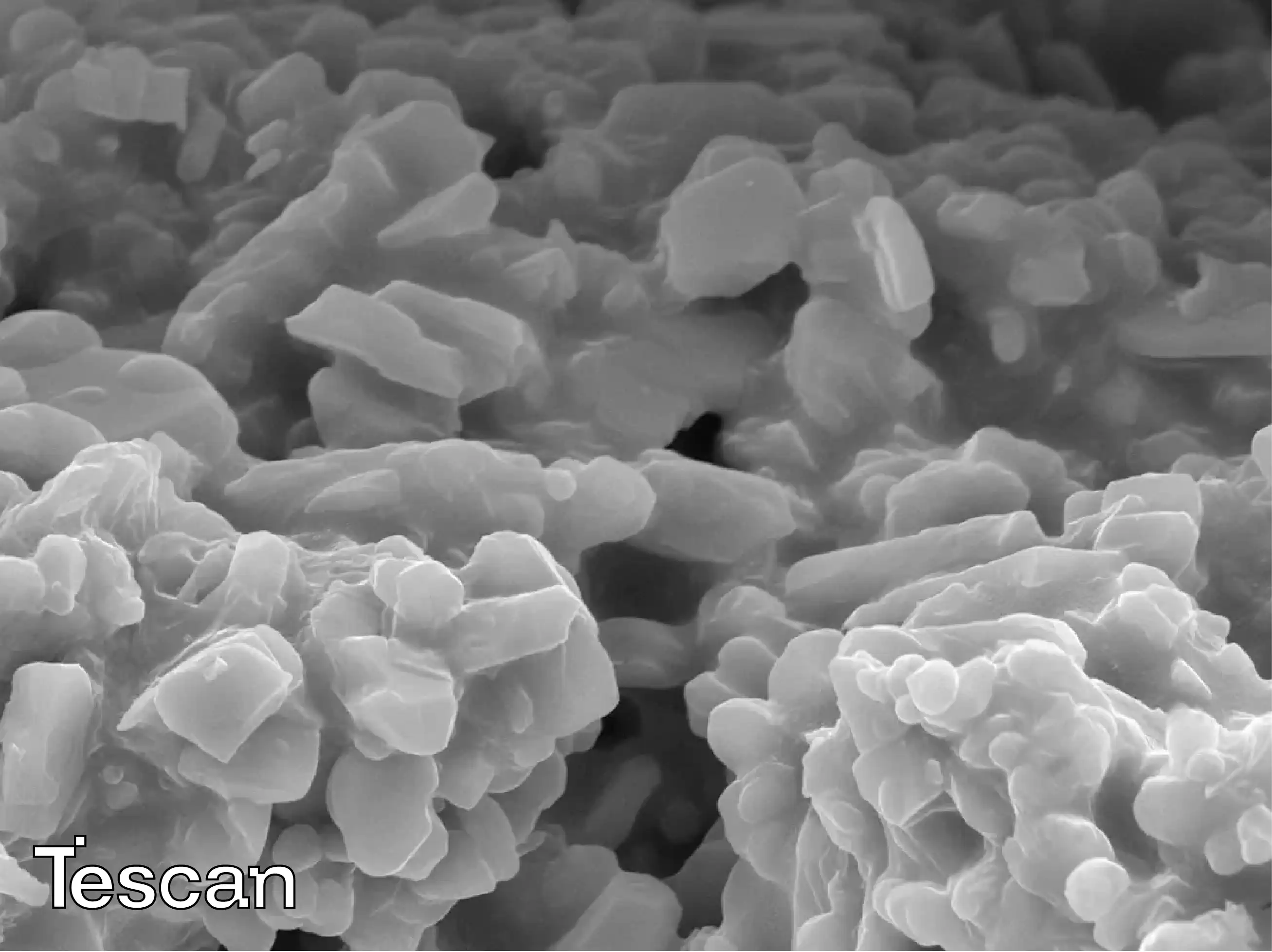
TEScan integrates advanced detector systems optimized for biological imaging. These detectors capture low-energy signals and fine topographical details, producing images with excellent contrast and depth perception. Enhanced signal processing ensures reliable data acquisition, even from soft or low-conductivity specimens. The result is a precise representation of biological textures essential for accurate structural interpretation.
Flexibility in Biological Research
TEScan SEM systems are designed to adapt to various life science workflows—from microbiological research to complex tissue studies. Flexible sample chambers accommodate a wide range of specimen sizes and holders, while modular configurations allow integration of correlative imaging tools. This adaptability supports both academic research and applied biomedical studies where workflow efficiency and precision are essential.
Precision, Reproducibility, and Stability
TEScan SEM provides high repeatability and imaging stability essential for quantitative biological research. Automated calibration, drift correction, and beam alignment ensure consistent imaging results across multiple sessions. This reproducibility is critical when comparing biological samples or monitoring changes over time in experimental studies.
Integration with Analytical Techniques
TEScan SEM systems integrate seamlessly with complementary analytical tools, expanding capabilities for life science applications. Coupling imaging with spectroscopy or 3D surface analysis enhances understanding of structural composition and functional relationships. This integrated approach supports comprehensive investigations into biological materials, bridging visualization and quantitative analysis.
Read more about Tescan products here
.webp)


.webp)
.webp)