


Home » Products » Confocal Microscopy and Imaging Systems » Confocal Microscopy » Andor Benchtop Confocal
Andor Benchtop Confocal
|
Fast to learn & time saving
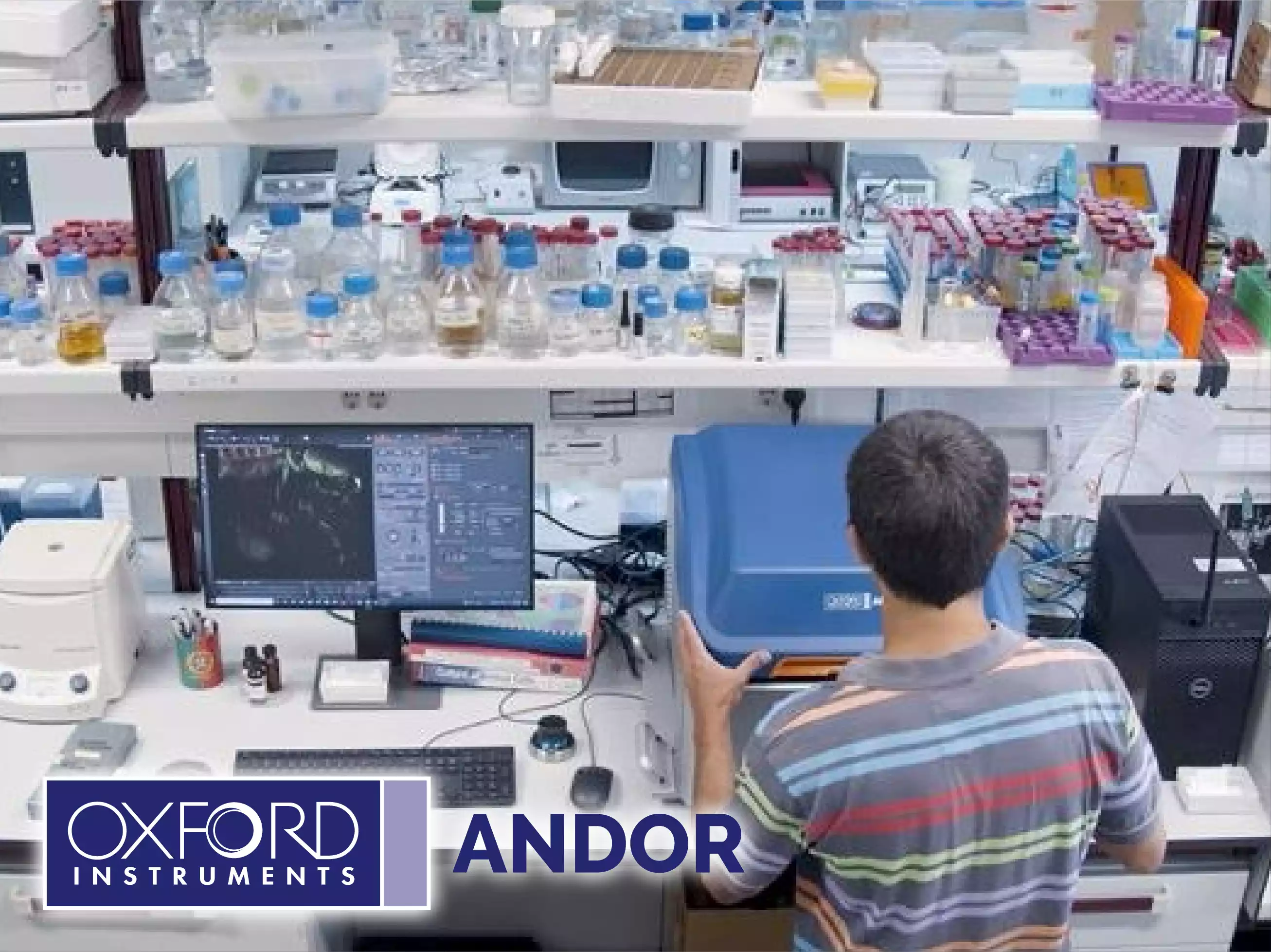
Andor Benchtop Confocal
BC43 captures images at least 10x faster than point scanning confocals, boosting productivity, yet maintaining full resolution. Image deeper with higher quality than solutions that rely on computational clearing or deconvolution alone. BC43 offers two transmitted light options: Brightfield for samples with inherent contrast like larger organisms, and Differential Phase Contrast (DPC), that can be applied for samples which deliver high and low contrast. You can even combine image modes for even greater imaging flexibility! For example, combine DPC with widefield or confocal imaging modalities.
Application Focus
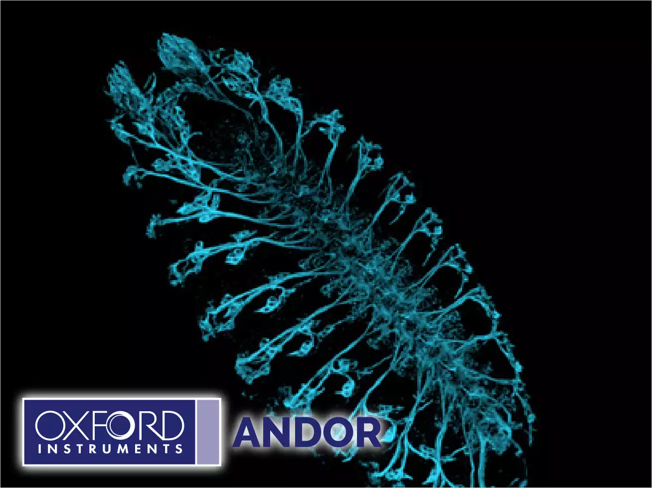
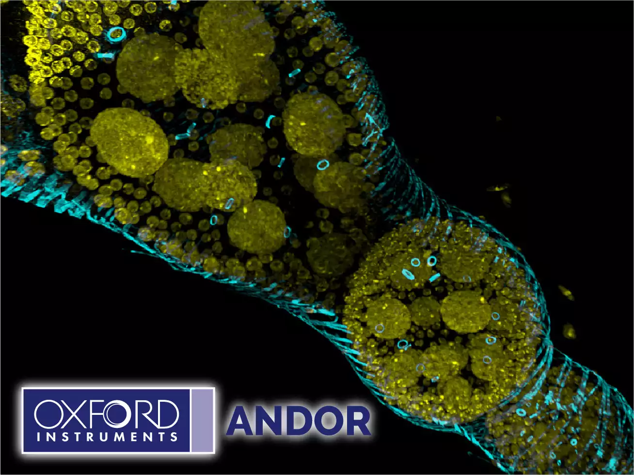
1- Developmental Biology
BC43 cuts through the challenges easily, spanning development from the first rounds of cell division to the fully developed organism. Use BC43 to image at depth, in gentle live imaging experiments of cells and tissues. Effortlessly acquire multiple Z stacks, multiple tiles in combination with time-lapse imaging. Extract sharp 2D images or instantly explore stunning 3D volumes in a fraction of the time you’re used to. BC43 delivers fast high-resolution imaging of developing model organisms (e.g. zebrafish and drosophila). Imaging deeper than conventional fluorescence microscopes and delivering a 10-fold more productive experience than a traditional confocal. No sacrificing sensitivity, resolution or 3D detail for speed, or to avoid bleaching.
BC43 features for development biology:
- Fast high resolution imaging.
- Image deep in both live and fixed samples.
- Montage & seamless stitching at any level of magnification.
Developmental Biology
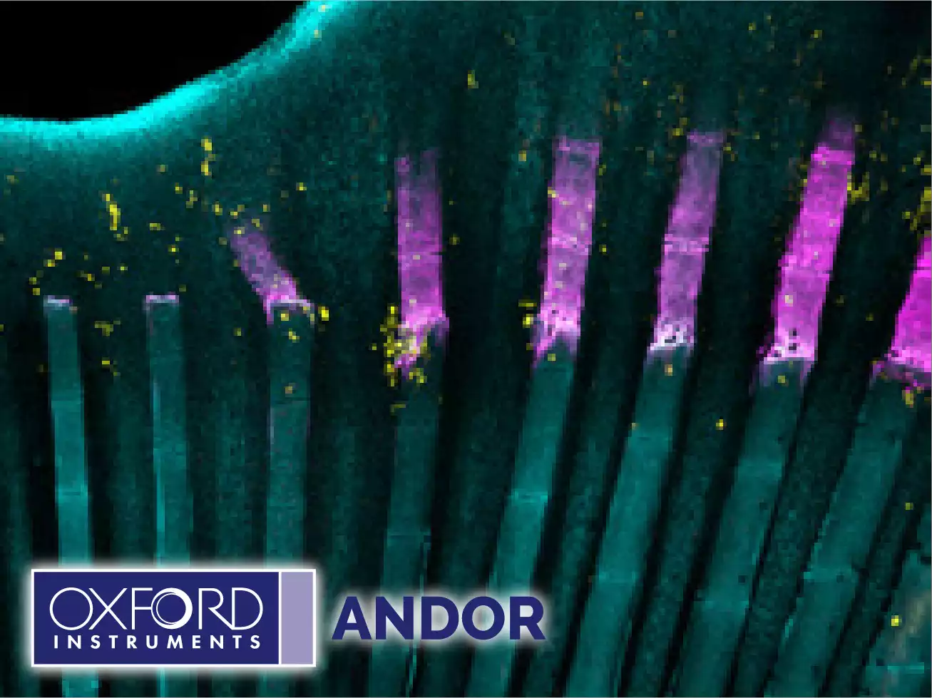
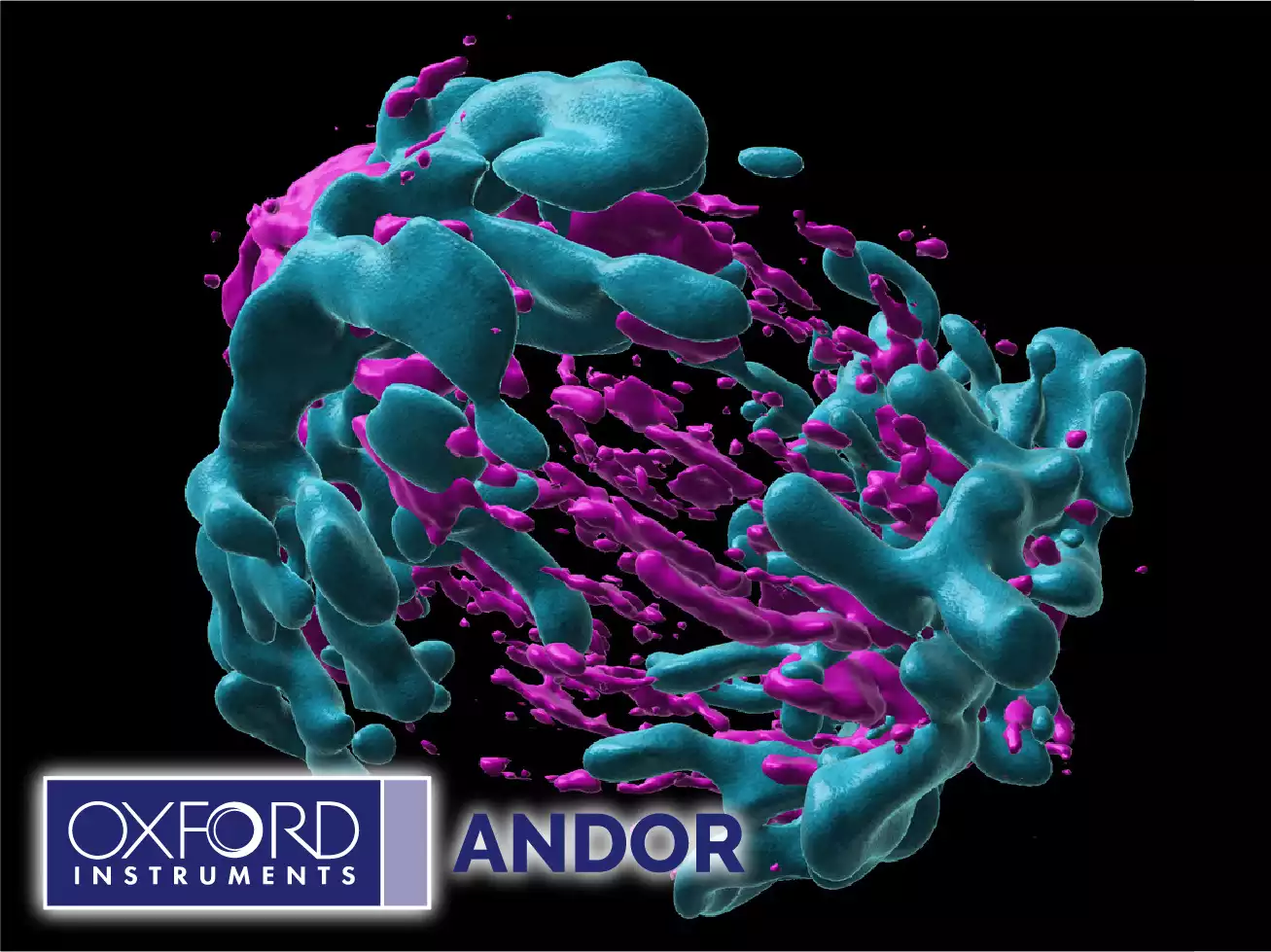
2- Cell Biology
Working closely with leading cell biologists we have carefully developed BC43 to meet the needs of a broad range of experiments. Reveal the detail inside cells from nm to mm within tissues and whole model organisms with BC43. Use BC43 in confocal mode to see detail hidden in the sample background or image in widefield to increase sensitivity and speed. Image fast dynamic events, such as microtubule dynamics, or study longer processes like cell cycle over 24 hours with no photobleaching or phototoxicity.
BC43 features for cell biology:
- Image long processes.
- Image fast dynamic events.
- No photobleaching or phototoxicity.
- nm to mm imaging capability.
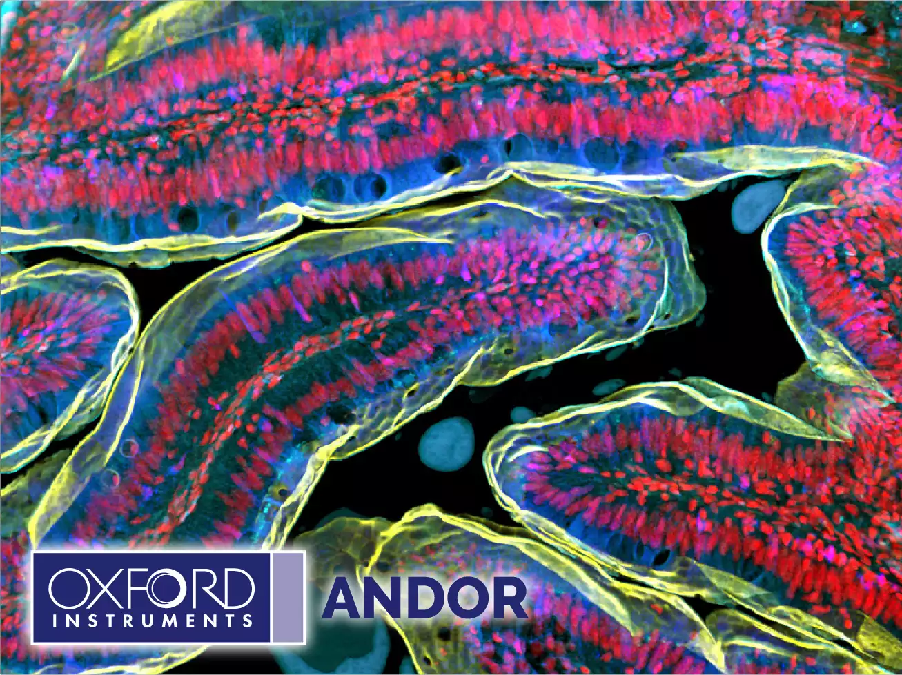
3- Tissue Imaging
Large area imaging needs to provide both cellular resolution and the full organ context. The advanced high-speed technology in BC43 means you no longer need to compromise. Large area tissue confocal imaging is now possible. Ten times faster than regular confocals. No sacrifices in resolution, or field of view. BC43 delivers
results fast, shortening the time to publication. Discover more in intact tissues, use cleared samples and BC43 in confocal mode to image even thicker samples. BC43 takes advantage of the working distance of modern objectives: imaging hundreds of microns at high magnifications, and beyond.
BC43 features for tissue imaging:
- Fast confocal and low light widefield imaging.
- Seamless large tissue imaging for fixed and live sample.
- Image from nm to mm
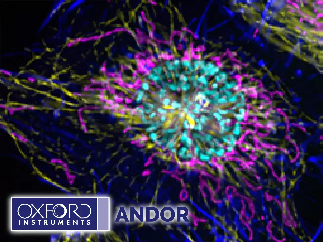
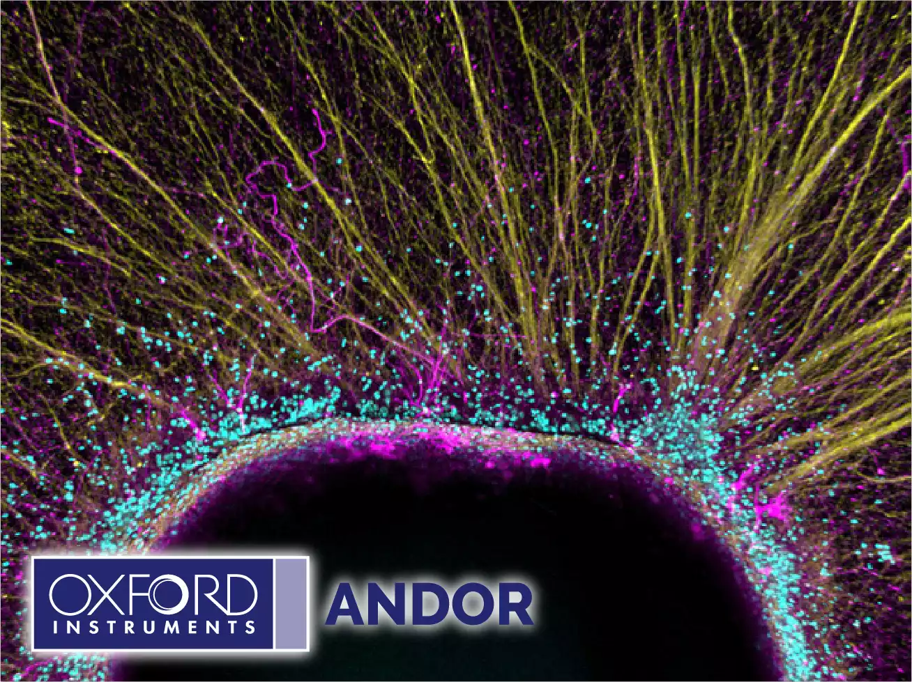
4- Neuroscience
BC43 is the perfect workhorse for neuroscience. Imaging experiments commonly require high magnification, for resolution, imaging of large areas to fully understand the architecture and connectivity of this complex tissue. The incredible confocal capture rate of BC43 dramatically reduces imaging time delivering results faster. Image both fixed and live samples, such as deep into cleared brain sections or developing organoids, covering the breadth of neuroscience microscopy needs.
Integrated Software Solutions
1- Fusion
BC43 has an integrated, easy-to-use, and accessible software interface that delivers high-end imaging. Users benefit from easy protocol set up for multidimensional experiments, such as one-click multi–position-montage and multiwell integration with an intuitive user interface and workflow for protocol set up. BC43 Fusion delivers real-time GPU-based deconvolution increasing the resolution of the image. Seamlessly integrated into the hardware, the in-line 3D stitching allows the full montage and visualisation of multiple tiles integrated into the context of the whole organism.
2- Imaris®
BC43 saves files in the Imaris IMS file format, permitting easy transfer of data into Imaris. Imaris for BC43 is included for isosurface rendering, high resolution snapshots, creation of multi-dimensional movies and downstream image editing. Additional application-specific modules of Imaris are available and include options for adding measurements suited for cell & developmental biologists, neuroscientists and many more disciplines within life sciences.
Read more about Andor Benchtop Confocal